Save flat 35% on Assignment
Read this academic research paper example & explore New Assignment Help USA's full range of academic writing services designed just for you—because great work deserves the perfect partner. Flip the page and let’s make it happen!
Question 1
The skeletal system is comprised of 206 bones and this system can be divided into two parts:
As a whole, one of the important functions of the skeleton system is the production of white and red blood cells in the bone marrow. Additionally, another important function of the skeleton muscle is:
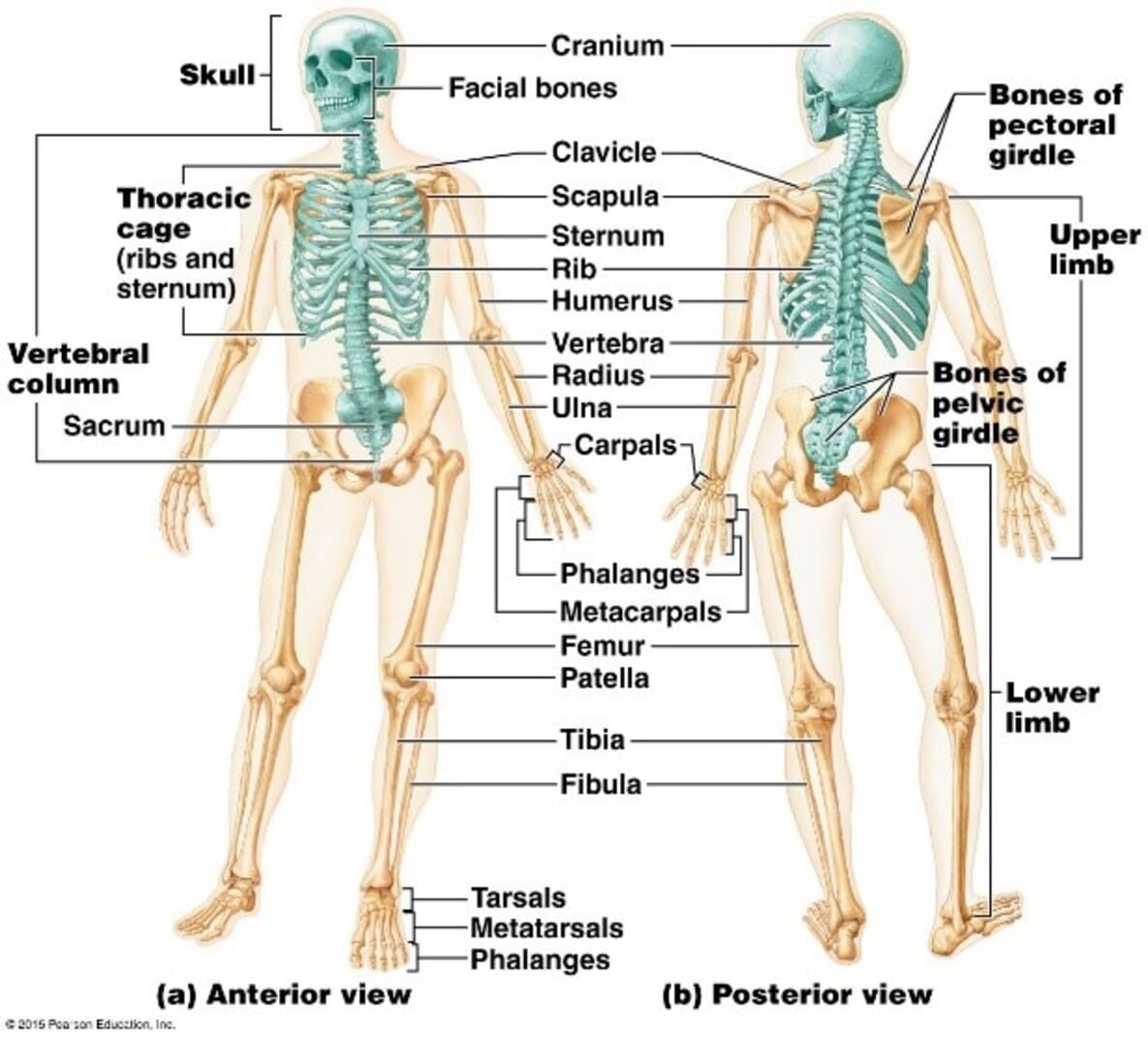
(Figure 1: Skeleton System of Human)
(Source: Pileggi, Parmar & Harper, 2021)
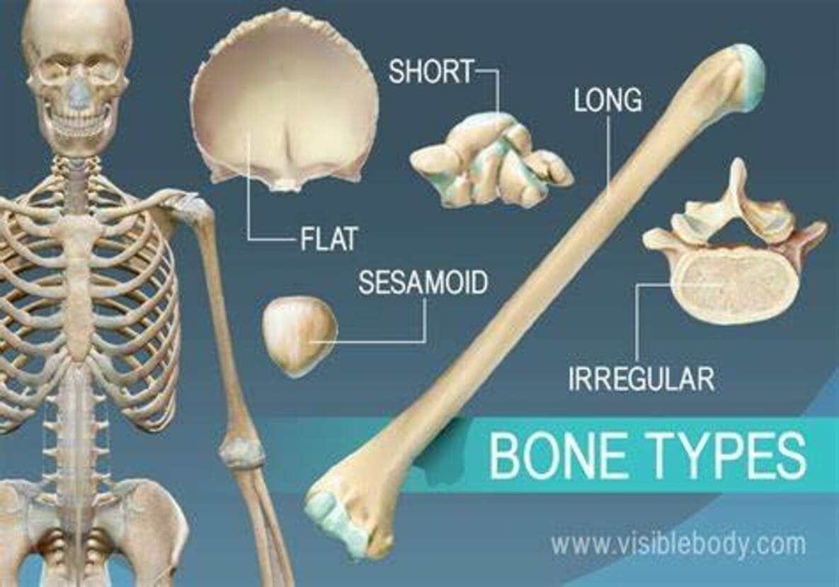
(Figure 2: Types of bones)
(Source: Pileggi, Parmar & Harper, 2021)
|
Type of joint |
Range of movement |
Example of type of joint in the body |
|
Fixed |
No Movement |
Skull- it is made up of fused bones |
|
Pivot |
Rotation around the central axis |
Neck and forearm |
|
Hinge |
Movement in one plane |
Elbow joints |
|
Ball and socket |
Rotate and turn in any direction |
Shoulder and hips |
Cartilage: Cartilage is flexible connective tissue that is made up of chondrocytes that give cartilage physical stability. The function of cartilage is to provide support and cushion the joints as it helps to maintain certain structures in the body (Pileggi, Parmar & Harper, 2021). Cartilage can be divided into three categories: elastic cartilage, fibrocartilage, and hyaline cartilage.
Ligaments: Ligaments are made up of connective tissues which connect one bone to another. The ligaments are made up of collagen fiber, which provides stability to the joints and prevents the dislocation of bones (Zhang, Liu & Zhang, 2021). Additionally, ligaments provide mobility to the joint. The knees are comprised of four ligaments which help knees to move side by side and backwards or upwards.
Tendons: Tendons are connected to muscle to bone. It is made up of collagen fiber, and within the tendon, three fibers are arranged in parallel bundles which give strength to the structure. The function of the tendon is to provide flexibility and withstand large stress (Mukund & Subramaniam, 2020).
Question 2
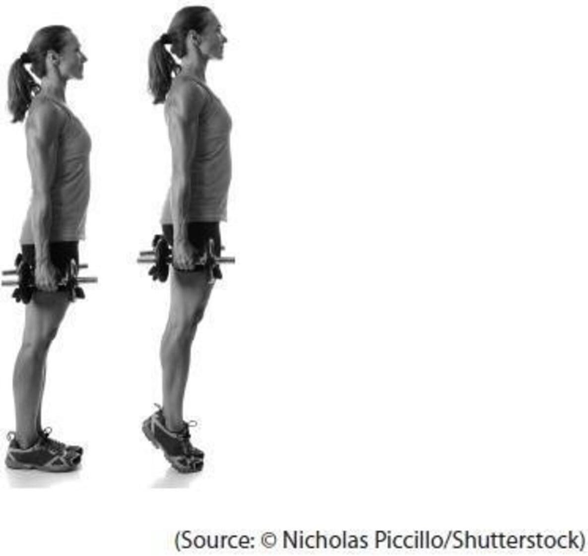
When a performer moves from standing onto her toes, it involves the use of a second-class lever system. In this lever system, the load is located between the fulcrum and the effort. The weight at the toes acts as the fulcrum, and the calf muscle supplies the upward effort and here the body weight acts as the downward load (Boyce & Schoenfeld, 2022). In this process, the gastrocnemius muscle plays an important role, in terms of contraction this muscle provides an upliftment energy of the heel, and the Achilles tendon pulls the heel to raise the entire body upward. This mechanism falls under the example of a second-class level mechanism in the human body.
Explain why this gives the athlete mechanical advantage. You may use diagrams to aid your explanation if you wish.
Considering the mechanism of the second-class lever system, it can be stated that this mechanism provides mechanical advantages to the athlete by allowing the calf muscle to generate greater force to uplift the body without much effort. Thus, the athlete can get flexibility and efficacy in the movement (Boyce & Schoenfeld, 2022). More specifically, it can be stated that the effort arm is larger than the load arm which provides mechanical advantages with relatively small efforts.
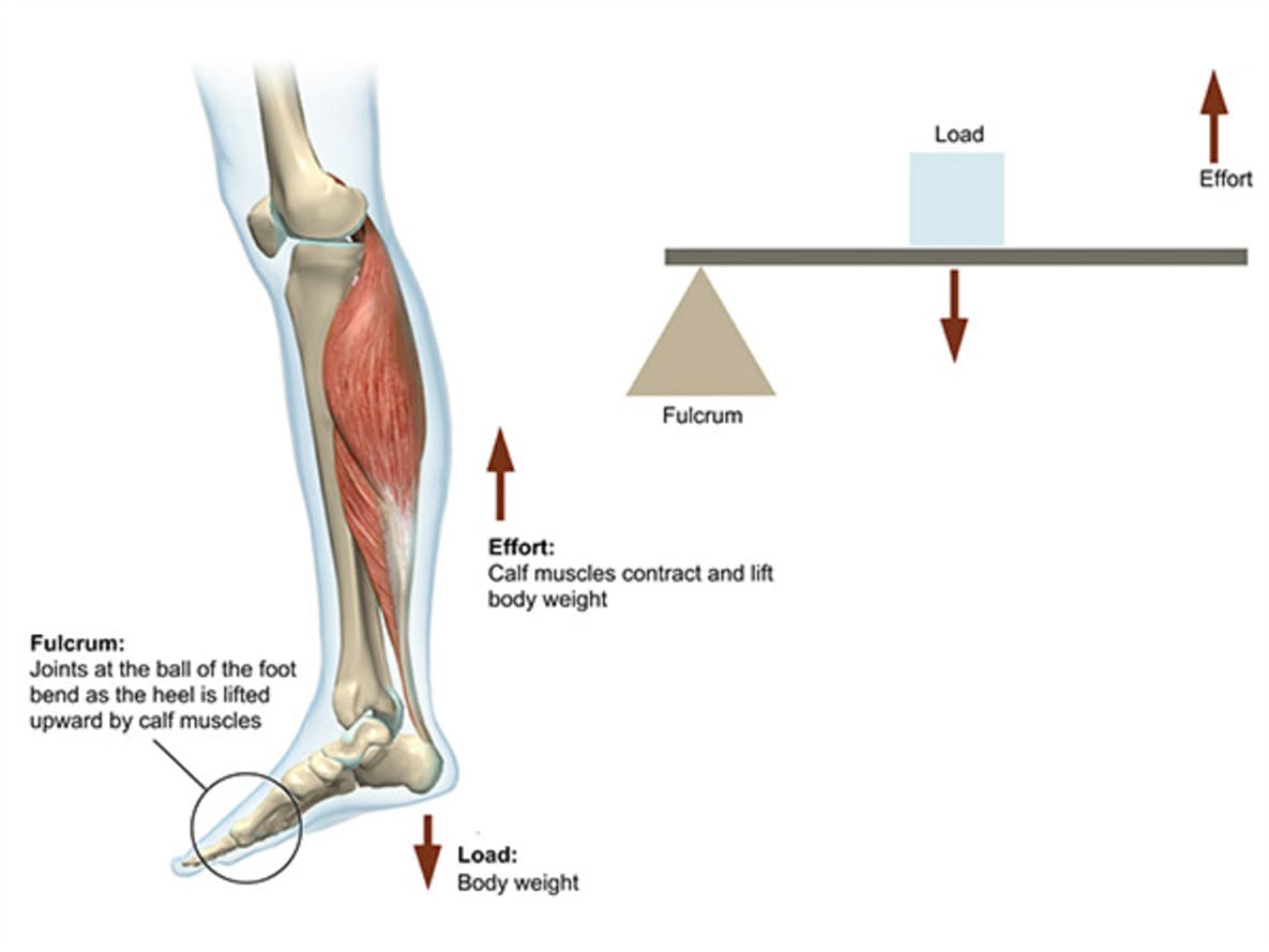
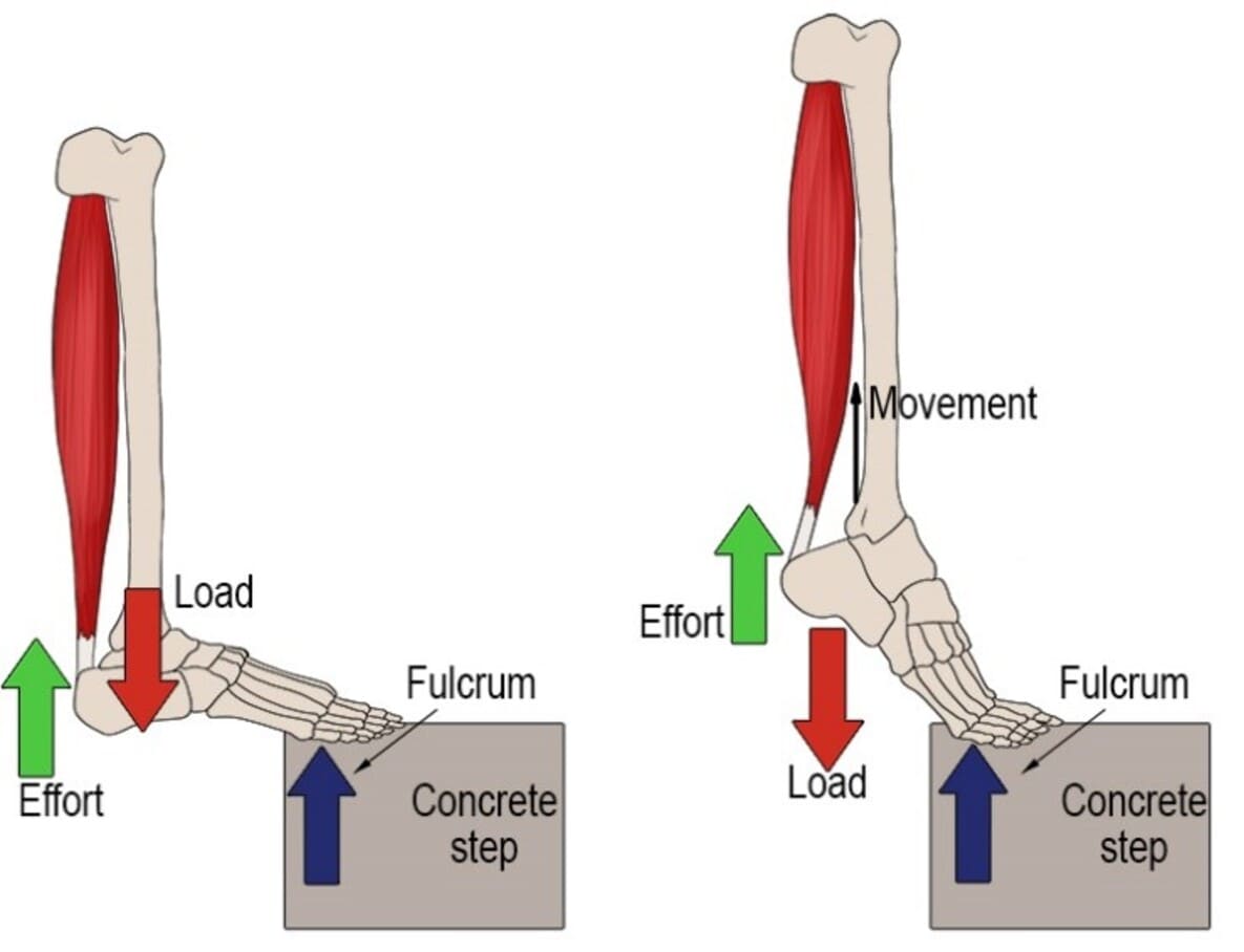
(Figure 3: Second-class lever)
(Source: Ulbrich et al. 2020)
Two examples of different classes of lever in athletic scenarios are:
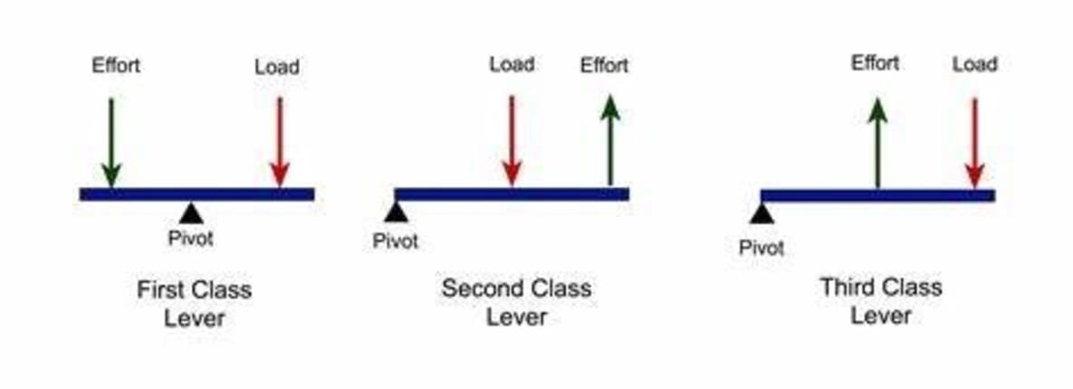
(Figure 4: Different types of levers)
(Source: Lattari et al. 2020)
Question 3
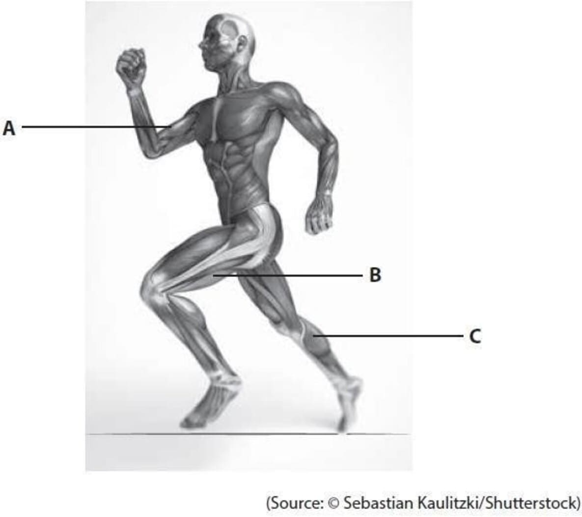
The image below shows the muscular system whilst running.
|
|
a) Muscle |
b) Role of Muscle |
|
A |
Biceps |
The location of the biceps is in the front of the upper arm. It is responsible for flexing the elbow and rotation of the forearm (Mukund & Subramaniam, 2020). It also stabilises the arm and shoulder while running. |
|
B |
Hamstrings |
It locates at the back of the thigh and between the knees and hips. It helps in hip extension and flexion of the knee. It also helps in thigh extension and helps to move the upper leg backward during running. |
|
C |
Calf muscles |
During running, the calf muscles are located at the back of the lower leg, and are responsible for plantar flexion, which helps to move the feet away from the field. It also helps to stabilise the ankle and foot while running (Glancy & Balaban, 2021) . |
C) Explain the structure of striated (skeletal muscle) and compare the muscle structure in areas of the body that move frequently to those that move less frequently.
Striated muscle is made up of long, and cylindrical muscle fibers and located in various parts of the body, including the neck, hands, feet, and tongue. The Striated muscle or skeletal muscle can be defined as the tissue that helps the muscle to be attached to the bone. It is voluntary and facilitates the voluntary movement of the bones (Powers et al. 2021). On the other hand, cardiac muscle is involuntary and found at the walls of the heart. Both the skeletal muscle and cardiac muscle have a striated appearance due to the presence of myofibrils (Morris, Zawieja & Moore Jr, 2021). Unlike the striated muscle, smooth muscles are involuntary in movement and its arrangement in the cell facilitates the relaxation and contraction of the muscle and provide elasticity.
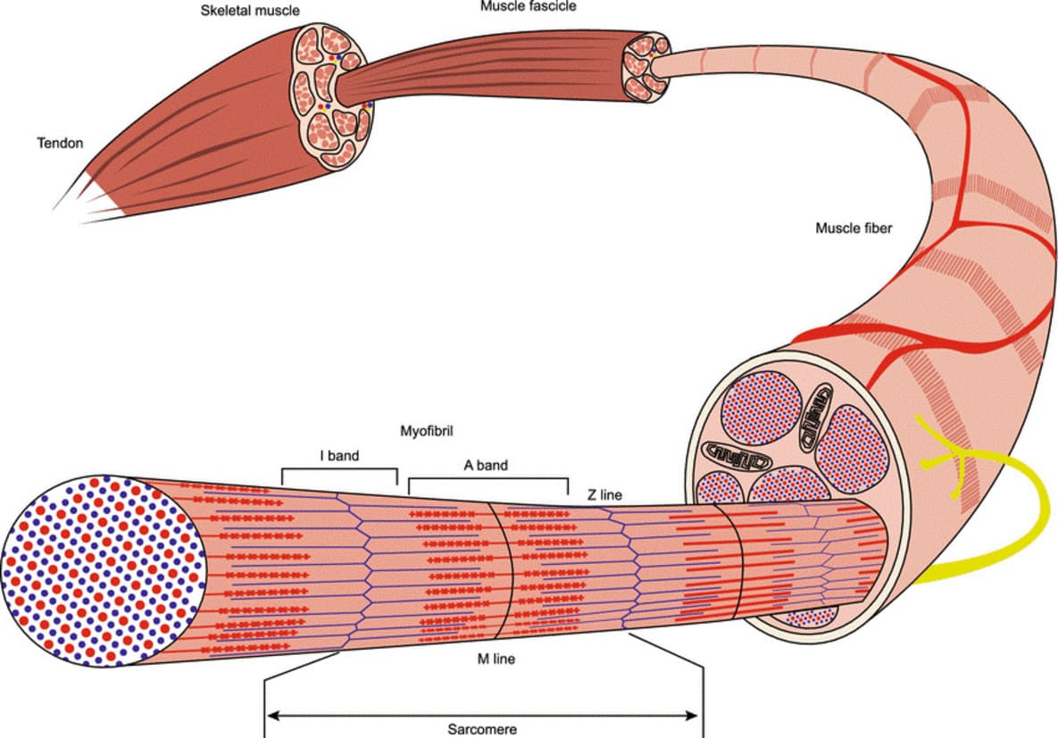
(Figure 5: Structure of Skeletal, Striated Muscle)
(Source: Sasikumari, 2020)
The sliding filament theory- this theory was propounded by A.F. Huxley and R. Niedegerke (1954). According to their observation, in one zone, the repeated sarcomere has formed an arrangement that can be referred to as “A Band”, which is constant in length during the contraction (Zhuge et al. 2020). The I band is rich in thinner filament which is made up of actin and myosin. According to the theory, sliding of actin and myosin can generate muscle tension, and actin located at the adjacent end of the sarcomere forms the “z-disc-like structure”, which can be called a Z-band (Saxton, Tariq & Bordoni, 2023). Considering this theory, it can be stated that in muscle contraction, the thin filament of action slides over the myosin, and this sliding facilitates the formation of a cross-bridge structure. In this structure, myosin heads undergo through a power stroke which causes the filament to slide over and cause shortening of the sarcomere and muscle fiber. During this time, the myosin filament is attached to the “M-line” at the center of the sarcomere whereas the actin filament is anchored to the “Z-line” at the end of the sarcomere and undergoes through conformational changes (Dong, Ma, & Guo, 2020).
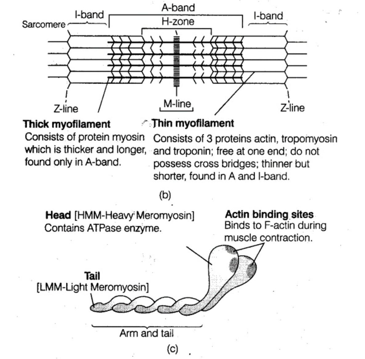
(Figure 6: Sliding Muscle Filament theory)
(Source: Dong, Ma & Guo, 2020)
Apart from the skeletal muscle, there is cardiac muscle and smooth muscle. The Cardiac muscle is accountable for the pumping in and out of the blood throughout the body. The movement of this muscle is involuntary and cardiac muscle is striated, and has a unique branching structure that can allow the cardiac muscle to pump in and out of the blood throughout the body simultaneously (Sasikumari, 2020).
On the other hand, Smooth muscles are located at the walls of the internal organs like the stomach, intestine, and blood vessels. Like Cardiac Muscle, Smooth muscle is also involuntary, but it is not striated. The shape of Smooth Muscle is spindle-like and arranged in a layer which can allow the muscle to contract and relax in a coordinated way (Saxton, Tariq & Bordoni, 2023).
Question 4
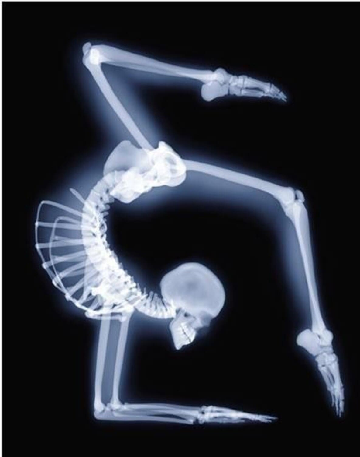
The process involving the growth and development of bone is known as ossification. The osteogenesis mainly occurs in two ways: intramembranous and endochondral.
The Intramembranous process facilitates the formation of primitive connective tissues. this process is also followed for the formation of flat bones in the skull, jaw, and clavicle (Salhotra et al. 2020). These bones are shaped flat during the formation and comprise with membrane-like layers of connective tissues that foster the blood supply. Therefore the processes involved are:
On the other hand, the endochondrial process uses the hyaline cartilage for the formation of long bones.
Training as a gymnast has positive as well as negative impacts on bone health. Gymnasium is related to the repetitive loading of bones which stimulates bone growth and density. On the other hand, a calorie-restricted diet can risk the onset of osteoporosis, a condition that can be defined by low bone density and the onset of fracture in the bone (Eckers, Fischer & Tscholl, 2020). On the other hand, calorie restriction can cause a reduction in the level of testosterone and estrone hormone in blood which play important roles in the formation and maintenance of bone. Therefore, to maintain bone health during the training period for gymnasts, it should be important to consume a balanced diet that is rich in nutrients for bone growth and development.
The first step of repairment of a fractured bone is the formation of the blood clot at the site of injury, which in turn can help to stabilise the bone and facilitate the mending processes. At that time, the body started the disposition of Calcium on the blood clots which turns into the soft callus. From this time, osteoblast and osteoclast are started to form (Bonazza et al. 2022). The osteoblast is the large cell that is responsible for the creation and laying down of minerals in bone, both at the time of the first formation of bone and later when the bone starts to change its structure throughout life (remodeling) (Makovitch & Eng, 2020). Osteoclast refers to the cells that are responsible for the breakdown and reabsorption of the bone tissues. The healing process takes several days or weeks or even several months based on the severity of the injuries or fractures. During this time, the bones started to reshape and strengthen through remodeling (Gissi et al. 2020). It involves the removal of the old bone tissues by the osteoclasts and the deposition of the new bone tissues by the osteoblasts.
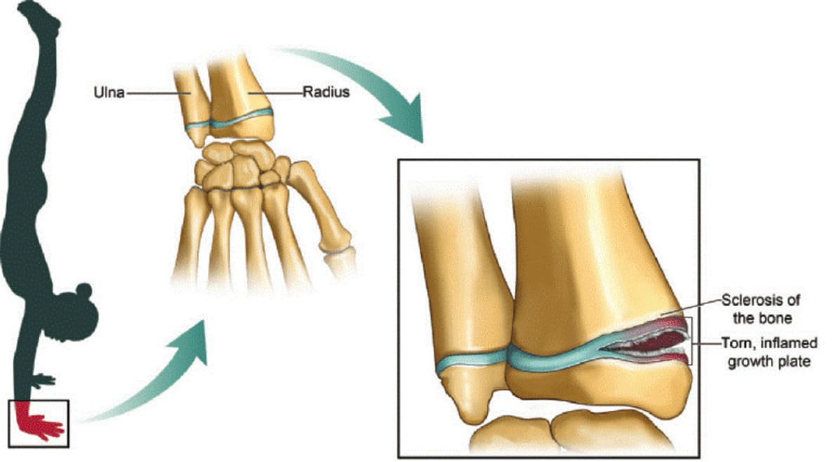
(Figure 8: Bone Injuries for Gymnasts)
(Source: Gissi et al. 2020)
In old age, the bone joints are affected by the sudden changes in connective tissues and cartilage. The cartilage inside the joint becomes thinner and components of the cartilage which can also be known as proteoglycans become changed, which makes the joints more prone to fracture or damage (Makovitch & Eng, 2020). Additionally, in old age, the connective tissues within the ligaments and tendons start to become rigid and brittle, which makes the joint stiff and fragmented. Loss of muscle, which can be termed as sarcopenia starts at the age of 30 and remains throughout life. In this process, the number of muscle tissues and the size of the muscle fibers started to decrease and cause loss of muscle mass and strength (Gissi et al. 2020). Some diseases like rheumatoid arthritis and osteoporosis cause inflammation at joints and reduce bone density, which in turn alleviates the fracture in the bone.
Bonazza, N. A., Saltzman, E. B., Wittstein, J. R., Richard, M. J., Kramer, W., & Riboh, J. C. (2022). Overuse elbow injuries in youth gymnasts. The American Journal of Sports Medicine, 50(2), 576-585. https://journals.sagepub.com/doi/abs/10.1177/03635465211000776
Boyce, L., & Schoenfeld, M. (2022). Strength Training for All Body Types: The Science of Lifting and Levers. Human Kinetics. https://books.google.com/books?hl=en&lr=&id=0-yaEAAAQBAJ&oi=fnd&pg=PR1&dq=lever+function+for+weight+lift+&ots=7UavngDfbc&sig=zl0YbGnutfnNaZoDUHNtL_aWXeY
Brunello, E., & Fusi, L. (2023). Regulating Striated Muscle Contraction: Through Thick and Thin. Annual Review of Physiology, 86.https://www.annualreviews.org/doi/abs/10.1146/annurev-physiol-042222-022728
Dong, R., Ma, P. X., & Guo, B. (2020). Conductive biomaterials for muscle tissue engineering. Biomaterials, 229, 119584. https://www.sciencedirect.com/science/article/pii/S0142961219306830
Eckers, F., Fischer, L., & Tscholl, P. M. (2020). Gymnastics. Injury and Health Risk Management in Sports: A Guide to Decision Making, 733-740. https://link.springer.com/chapter/10.1007/978-3-662-60752-7_111
Gissi, C., Radeghieri, A., Antonetti Lamorgese Passeri, C., Gallorini, M., Calciano, L., Oliva, F., ... & Berardi, A. C. (2020). Extracellular vesicles from rat-bone-marrow mesenchymal stromal/stem cells improve tendon repair in rat Achilles tendon injury model in dose-dependent manner: A pilot study. PLoS One, 15(3), e0229914. https://journals.plos.org/plosone/article?id=10.1371/journal.pone.0229914
Glancy, B., & Balaban, R. S. (2021). Energy metabolism design of the striated muscle cell. Physiological Reviews, 101(4), 1561-1607. https://journals.physiology.org/doi/abs/10.1152/physrev.00040.2020
Kim, K., Reid, B. A., Casey, C. A., Bender, B. E., Ro, B., Song, Q., ... & Roseguini, B. T. (2020). Effects of repeated local heat therapy on skeletal muscle structure and function in humans. Journal of Applied Physiology, 128(3), 483-492. https://journals.physiology.org/doi/abs/10.1152/japplphysiol.00701.2019
Lattari, E., Rosa Filho, B. J., Junior, S. J. F., Murillo-Rodriguez, E., Rocha, N., Machado, S., & Neto, G. A. M. (2020). Effects on volume load and ratings of perceived exertion in individuals' advanced weight training after transcranial direct current stimulation. The Journal of Strength & Conditioning Research, 34(1), 89-96. https://journals.lww.com/nsca-jscr/fulltext/2020/01000/effects_on_volume_load_and_ratings_of_perceived.11.aspx
Makovitch, S., & Eng, C. (2020). Spine injuries in gymnasts. Gymnastics Medicine: Evaluation, Management and Rehabilitation, 135-176. https://link.springer.com/chapter/10.1007/978-3-030-26288-4_8
Miyazaki, Y., Ichimura, A., Kitayama, R., Okamoto, N., Yasue, T., Liu, F., ... & Takeshima, H. (2022). C-type natriuretic peptide facilitates autonomic Ca2+ entry in growth plate chondrocytes for stimulating bone growth. Elife, 11, e71931. https://elifesciences.org/articles/71931
Morris, C. J., Zawieja, D. C., & Moore Jr, J. E. (2021). A multiscale sliding filament model of lymphatic muscle pumping. Biomechanics and Modeling in Mechanobiology, 20(6), 2179-2202.https://link.springer.com/article/10.1007/s10237-021-01501-0
Mukund, K., & Subramaniam, S. (2020). Skeletal muscle: A review of molecular structure and function, in health and disease. Wiley Interdisciplinary Reviews: Systems Biology and Medicine, 12(1), e1462. https://wires.onlinelibrary.wiley.com/doi/abs/10.1002/wsbm.1462
Mukund, K., & Subramaniam, S. (2020). Skeletal muscle: A review of molecular structure and function, in health and disease. Wiley Interdisciplinary Reviews: Systems Biology and Medicine, 12(1), e1462.https://wires.onlinelibrary.wiley.com/doi/abs/10.1002/wsbm.1462
Pileggi, C. A., Parmar, G., & Harper, M. E. (2021). The lifecycle of skeletal muscle mitochondria in obesity. Obesity Reviews, 22(5), e13164.https://onlinelibrary.wiley.com/doi/abs/10.1111/obr.13164
Powers, J. D., Malingen, S. A., Regnier, M., & Daniel, T. L. (2021). The sliding filament theory since Andrew Huxley: multiscale and multidisciplinary muscle research. Annual review of biophysics, 50, 373-400. https://www.annualreviews.org/doi/abs/10.1146/annurev-biophys-110320-062613
Salhotra, A., Shah, H. N., Levi, B., & Longaker, M. T. (2020). Mechanisms of bone development and repair. Nature reviews Molecular cell biology, 21(11), 696-711. https://www.nature.com/articles/s41580-020-00279-w
Sasikumari, O. (2020). Defects in the Existing Theory of Skeletal Muscle Contraction and Postulation of a New Theory. European Journal of Medical and Health Sciences, 2(6). https://www.ej-med.org/index.php/ejmed/article/view/573
Saxton, A., Tariq, M. A., & Bordoni, B. (2023). Anatomy, thorax, cardiac muscle. In StatPearls [Internet]. StatPearls Publishing. https://www.ncbi.nlm.nih.gov/books/NBK535355/
Ulbrich, J., Swader, R., Petry, G., Cox, B. L., Greene, R. L., Eliceiri, K. W., & Radwin, R. G. (2020). A syringe adapter for reduced muscular strain and fatigue. Applied Ergonomics, 85, 103061. https://www.sciencedirect.com/science/article/pii/S000368702030017X
Yamamoto, M., Sakiyama, K., Kitamura, K., Yamamoto, Y., Takagi, T., Sekiya, S., ... & Abe, S. (2022). Development and regeneration of muscle, tendon, and myotendinous junctions in striated skeletal muscle. International journal of molecular sciences, 23(6), https://www.mdpi.com/1422-0067/23/6/3006.
Zhang, W., Liu, Y., & Zhang, H. (2021). Extracellular matrix: An important regulator of cell functions and skeletal muscle development. Cell & bioscience, 11, 1-13.https://link.springer.com/article/10.1186/s13578-021-00579-4
Zhuge, Y., Zhang, J., Qian, F., Wen, Z., Niu, C., Xu, K., ... & Jia, C. (2020). Role of smooth muscle cells in Cardiovascular Disease. International Journal of Biological Sciences, 16(14), 2741. https://www.ncbi.nlm.nih.gov/pmc/articles/PMC7586427/
